MRI Vs. CT Scan After a Car Accident: What Is Best?


Every article on this site is researched by our internal team, reviewed for legal accuracy against current Texas law, and held to State Bar of Texas advertising standards before publication. We do not publish content that overstates outcomes or makes promises about results.
Learn more about our
editorial standards .
Key Takeaways
- CT scans find fractures and bleeding; MRI finds disc, ligament, and nerve damage.
- A normal emergency room CT scan does not rule out a soft-tissue or delayed injury.
- Texas gives you two years to sue; document imaging dates and your doctor's rationale.
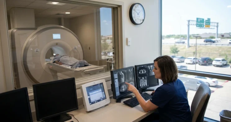
You walked out of the emergency room after a wreck on I-35 holding discharge papers that said your CT scan was normal. Three days later your neck still will not turn, and your fingers have started tingling.
A CT scan and an MRI are not better and worse versions of the same test. They answer different clinical questions.
A CT scan is the fast first look for broken bones and acute bleeding. An MRI is the more sensitive test for discs, ligaments, soft tissue, and the spinal cord.
So a clear CT scan does not mean you were not hurt. It means the scan you got did not find the kind of injury it was built to find.
What a CT Scan & an MRI Each Actually Show After a Car Accident
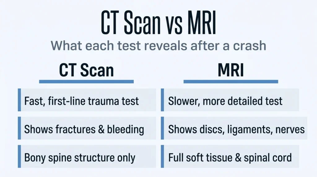
The MRI versus CT scan question after a car accident is not about scan quality. Emergency staff reach for CT first because they are looking for the injuries that can kill you in the next hour.
What a CT Scan Shows
A CT scan combines multiple x-ray images to show bone, soft tissue, and blood vessels in a single exam. It is fast, widely available, and in an emergency it can reveal internal injuries and bleeding quickly enough to help save lives.
That speed comes with tradeoffs. For the spine, a CT scan shows the bony vertebral structure clearly and can show disc position to some degree, but it does not consistently show enough detail to properly assess the spinal cord.
The same pattern holds for the head. A head CT detects bleeding, brain injury, and skull fractures quickly, but the precise details of soft tissue are less visible than on MR imaging.
This is why the American College of Radiology Appropriateness Criteria rates CT of the cervical spine without intravenous contrast as usually appropriate for initial imaging of blunt trauma patients aged 16 and older who meet standard screening criteria. CT is the right first test. It was never meant to be the last one.
What an MRI Shows
An MRI distinguishes abnormal tissue from normal tissue far more accurately than x-ray or CT, and it is described as the best available method to evaluate ligament injuries. It can show very small tears in tendons, ligaments, and muscles that do not appear on x-rays or CT scans.
For the spine, MRI is currently the most sensitive imaging test available, identifying damage to bone,
disc, ligament, or spinal cord. One balancing exception is worth knowing: some vertebral fractures are still better detected by CT.
The American College of Radiology criteria reflect that same split. Once ligament, spinal cord, or nerve root injury is suspected or confirmed, MRI of the area of interest becomes the usually appropriate next study after CT.
Why a Normal Emergency Room CT Scan Does Not Rule Out an Injury
A clear CT scan tells you something real and something limited. At the moment of that scan, the radiologist did not see a fracture or acute bleeding. It does not tell you that your discs, ligaments, and nerves came through the crash intact.
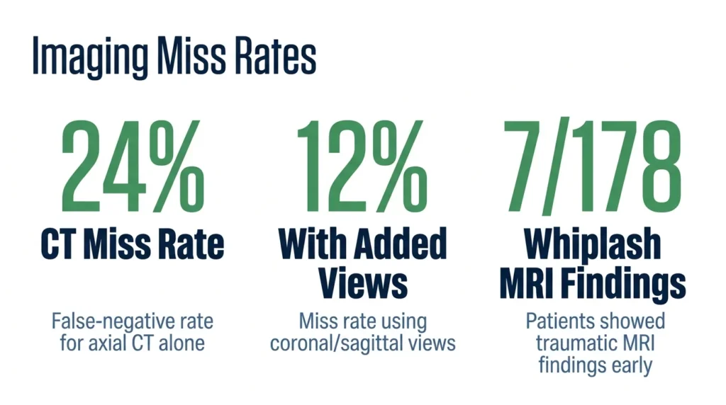
Emergency imaging can also miss injuries it is designed to catch. A National Institutes of Health case report describes a trauma patient whose initial head CT was read as normal. Three weeks later, with worsening symptoms, a repeat scan revealed a large subdural hematoma that required emergency surgery.
The literature reviewed in that report found a false-negative rate of roughly 24 percent for axial CT alone in acute subdural hematoma, dropping to about 12 percent once coronal and sagittal reformatted views were added. Those figures describe subdural hematoma specifically. The broader point they support is that normal emergency imaging does not exclude a bleed, particularly when symptoms persist or worsen.
Whiplash-type injuries sit in a different blind spot. They usually affect muscles, ligaments, and tendons rather than bone, which is exactly the tissue a bone-and-bleed scan handles least well.
Be careful about the opposite assumption too. A prospective one-year study of one hundred seventy-eight whiplash patients found traumatic findings on early cervical MRI in only seven of them, and those findings were not associated with outcome at three or 12 months. The authors concluded that absence of MRI findings does not indicate absence of injury.
Read those two studies together and you get the honest answer. A normal scan of either kind is not proof that nothing is wrong, and a later MRI gets ordered because it is the appropriate next step when symptoms continue, not because it is certain to produce a picture of your pain.
How an Imaging Gap Becomes a Fight Over What Caused Your Injury
Insurance adjusters read your imaging timeline very differently than your doctor does. An adjuster treats a clear CT scan as proof of no injury, and treats the weeks before anyone ordered an MRI as room to argue that something other than the crash caused what the MRI eventually found.
That is the whole move. A documented gap between the collision and the imaging that identified your injury gives the insurer an opening to offer an alternative explanation, which shifts the dispute from what your claim is worth to whether the wreck caused it at all.
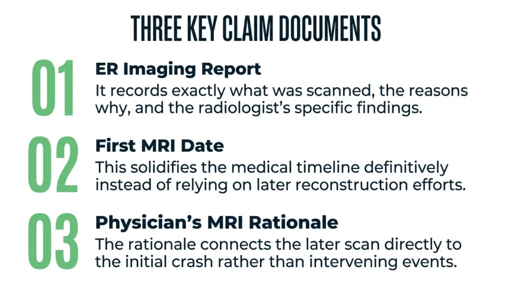
You do not beat that argument by disagreeing with it. You beat it with records. In a Texas car accident claim, three documents carry most of the weight:
- The emergency imaging order and report, which show what was scanned, why it was ordered, and what the radiologist actually wrote
- The date of your first MRI, which fixes the medical timeline instead of leaving it to be reconstructed later
- Your treating physician’s documented rationale for ordering that MRI, which is what ties the later scan back to the crash rather than to some intervening event
The third item does the most work. A physician’s written reason for escalating imaging turns what looks like a gap in time into a clinical sequence that follows your symptoms.
Timing matters in another way. Texas Civil Practice and Remedies Code Section 16.003 requires a person to bring suit for personal injury not later than two years after the cause of action accrues, which for a car accident is ordinarily the date of the collision. Symptoms that surface weeks later do not reset that clock.
What to Do if You Still Have Symptoms After a Clear CT Scan
If pain, numbness, or neurological symptoms are still with you after a normal CT scan, the next moves are straightforward:
- Tell your treating physician about every ongoing or worsening symptom. Persistent symptoms are what appropriately trigger an MRI referral under the clinical criteria.
- Ask your physician to document the specific reason for ordering an MRI, including persistent symptoms and any suspected ligament, disc, or nerve involvement.
- Keep the emergency imaging order and report, every follow-up visit record, and the date of any MRI together in one place.
- Do not read a clear CT scan at the end of your claim. It means the CT found no fracture and no acute bleed. It does not mean nothing is wrong.
- Talk to a personal injury attorney early, so your medical timeline is documented while it is happening rather than rebuilt from memory months later.
Talk to an Attorney About Your Imaging and Your Claim
A normal CT scan is not the end of your story, and the weeks between your emergency room visit and your first MRI should not quietly decide your claim.
Angel Reyes & Associates has guided injured Texans through the legal process for over 30 years, with board-certified personal injury attorneys and investigators on the team. They can review your records, your imaging timeline, and your options while the details are still fresh.
Contact Angel Reyes & Associates for a free consultation.
Past results do not guarantee future outcomes.
MRI & CT Scan FAQs
Can a car accident cause a herniated disc even if the first CT scan was normal?
Yes. A car accident can cause or aggravate a herniated disc, and a normal CT scan does not reliably rule out disc injury. A physician may recommend an MRI when symptoms suggest disc damage or nerve compression.
What symptoms after a car accident may indicate a spinal cord or nerve injury?
Numbness, tingling, weakness, loss of coordination, or pain that travels into the arms or legs may indicate nerve involvement and should be evaluated by a medical professional. New or worsening weakness, difficulty walking, or loss of bladder or bowel control requires urgent medical attention.
Can an MRI show an old injury versus a new injury from a car accident?
An MRI can reveal findings that help a physician assess the nature and possible timing of an injury, but it generally cannot establish the exact date an injury occurred by itself. Medical records, prior imaging, symptom history, and the treating physician’s interpretation may help distinguish a preexisting condition from a new or aggravated injury.
Does health insurance cover an MRI after a car accident in Texas?
Health insurance may cover a medically necessary MRI, but coverage depends on the policy, medical necessity requirements, and whether prior authorization is required. If another driver caused the accident, the costs may also become part of a personal injury claim, depending on liability and the available insurance coverage.
Can you recover compensation for a car accident injury if your MRI was delayed?
A delayed MRI does not automatically prevent recovery in a Texas personal injury claim. The significance of the delay depends on the medical evidence, the cause of the injury, the treatment timeline, and whether the available records support that the accident caused or aggravated the condition.
About the Authors

Angel Reyes
Writer
Angel L. Reyes, III is the Founder and Managing Partner of Angel Reyes & Associates. A nationally recognized trial attorney and business leader, Angel founded the firm in 1993, which has gone on to help over 70,000 injury victims recover more than $1 billion. Named to the Texas Super Lawyers list annually since 2008, Angel brings Wall Street experience, Ivy League education, and a deep commitment to client advocacy. He is a frequent legal commentator and published author with extensive contributions to legal scholarship, trial strategy, and consumer protection.

Graham Griffin
Editor
Graham Griffin is the Web & Content Manager at Angel Reyes & Associates, where he oversees content strategy, AI-powered workflows, a...

Angel Reyes
Reviewer
Angel L. Reyes, III is the Founder and Managing Partner of Angel Reyes & Associates. A nationally recognized trial attorney and business...